Northampton Advanced Imaging Facility (NAIF)
The Northampton Advanced Imaging Facility (NAIF) is the University of Northampton’s core facility for advanced imaging and flow cytometry. A wide range of equipment, training and services are available for both internal users and external users from other academic institutions and industry.
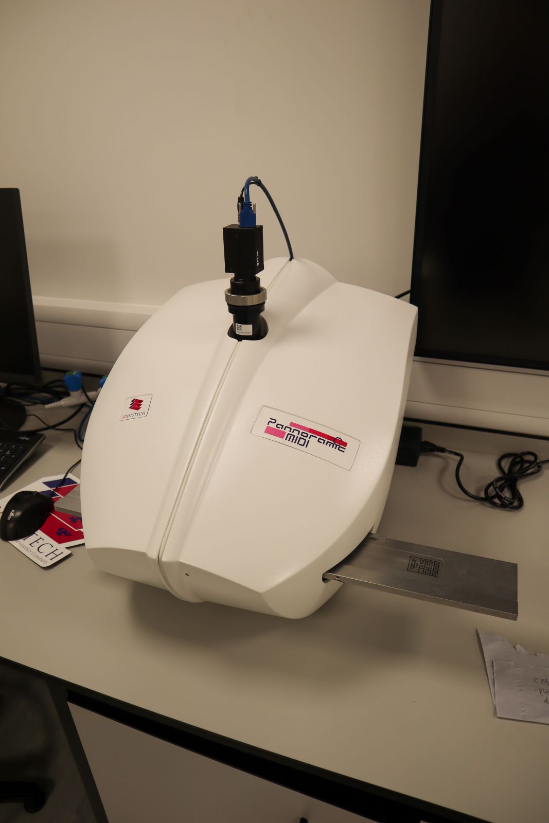
The features of our Epredia Pannoramic MIDI ll slide scanner include automatic loading, previewing and barcode reading.
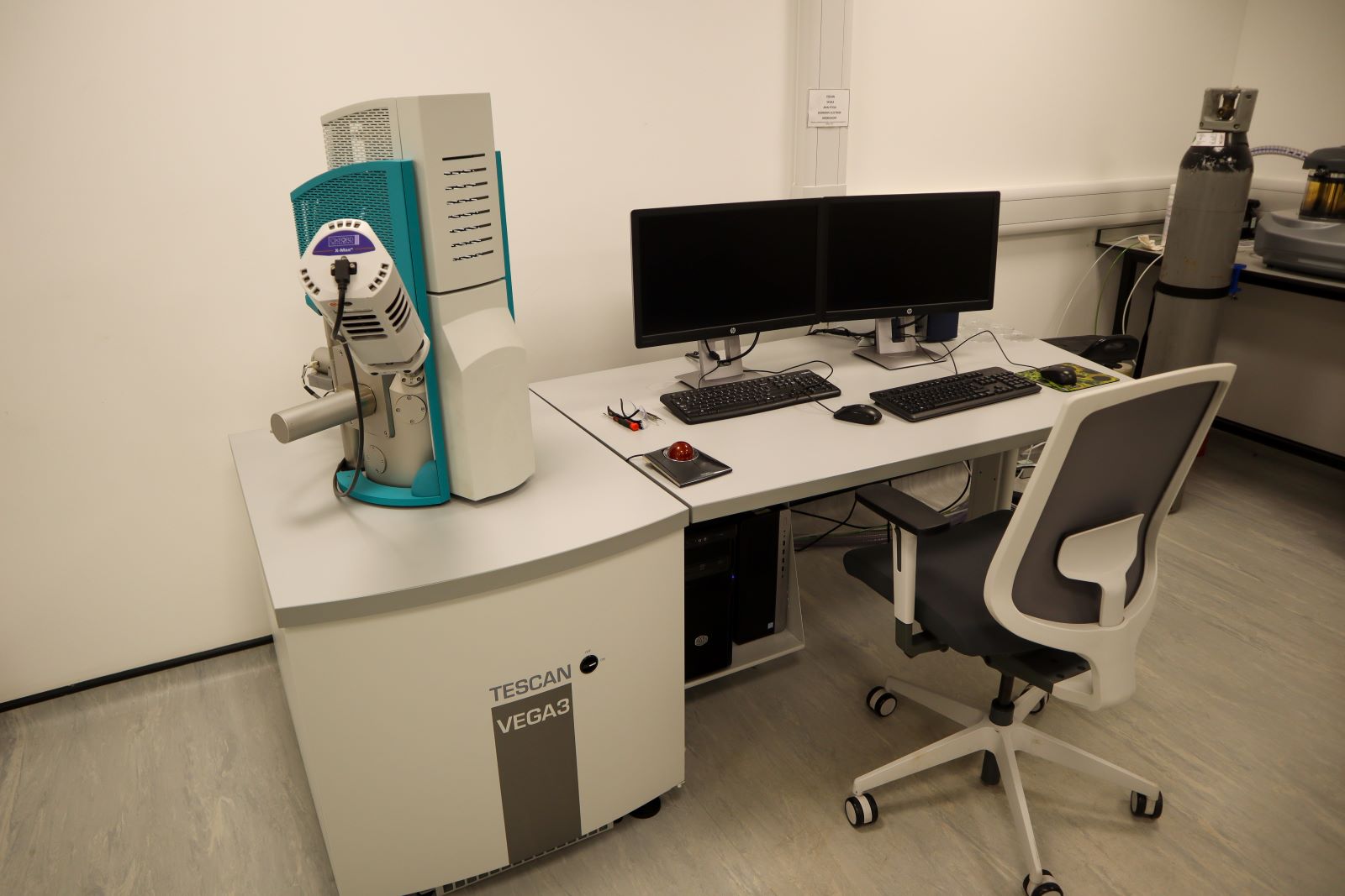
Our Scanning Electron Microscopy (SEM) is capable of extremely high magnifications far beyond what can be achieved with a traditional light microscope.
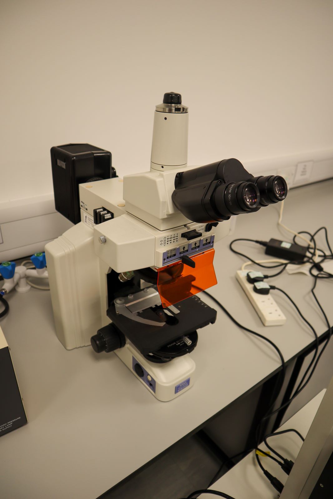
At Northampton Advanced Imaging Facility, we have one inverted and three upright epifluorescent microscopes available for quick and easy imaging of fluorescent samples.
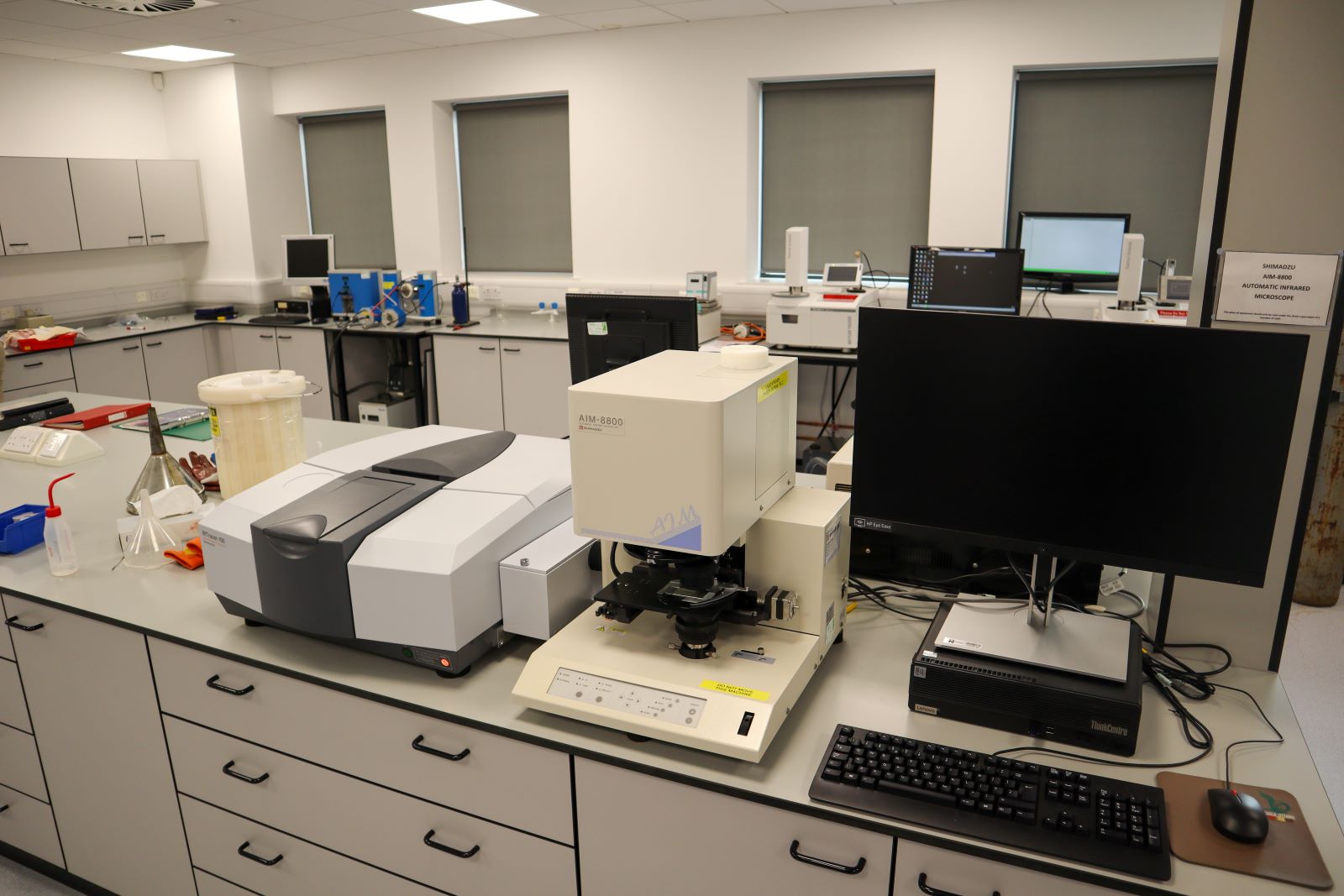
Our FTIR (Fourier transform infrared) spectroscopy machine is also combined with an infrared microscope which can provide high magnification infrared images allowing for further detailed analysis.
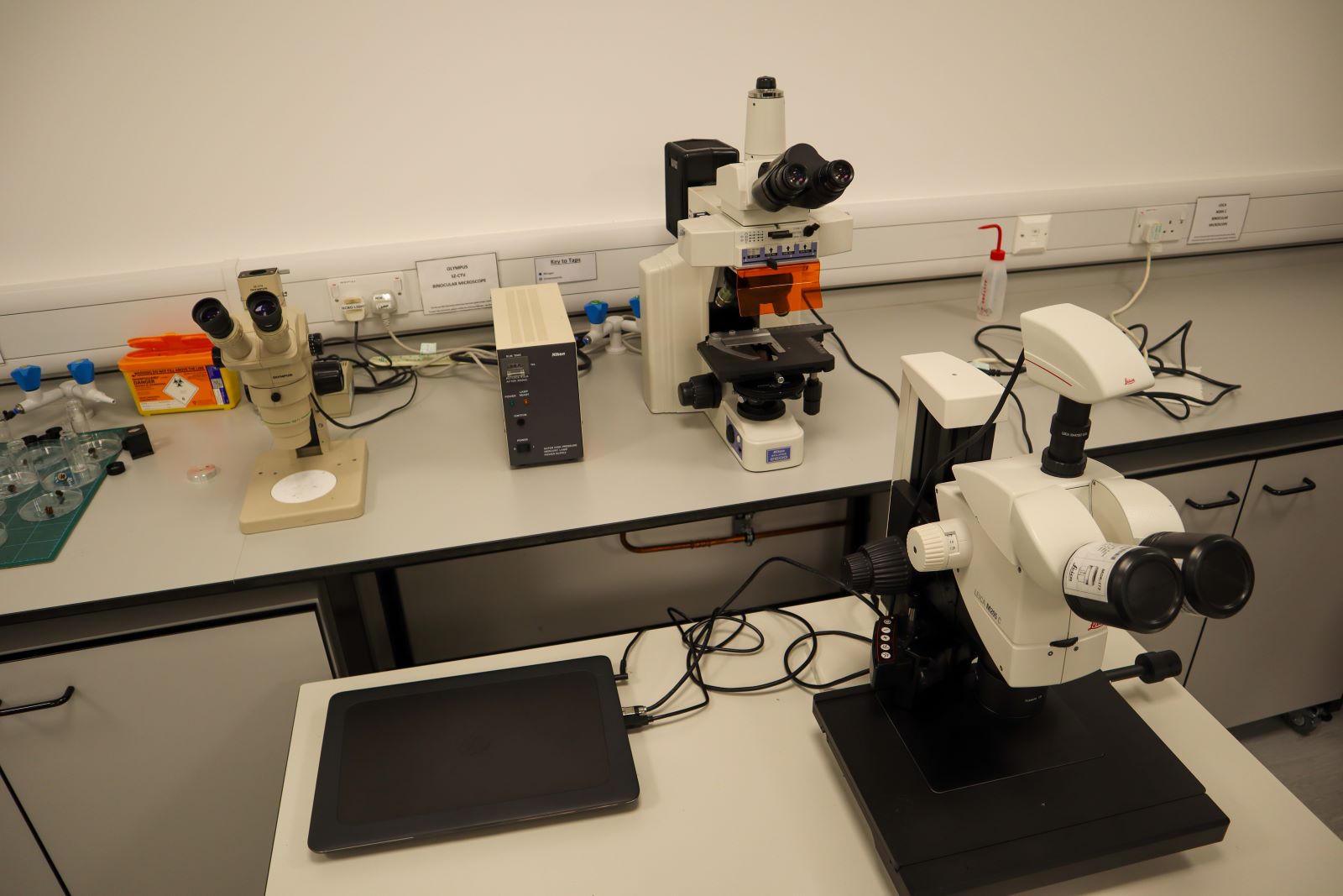
We have a range of digital and optical microscopes and software for the imaging and analysis of a variety of materials.
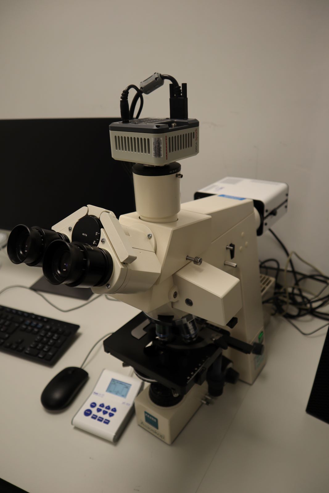
Our fluorescence microscopes are all equipped with digital cameras and user-friendly image processing software for quantitative analysis.

Our CytoFLEX flow cytometry platform is equipped with three active lasers and nine channels for fluorescent detection across 13 repositionable bandpass filters
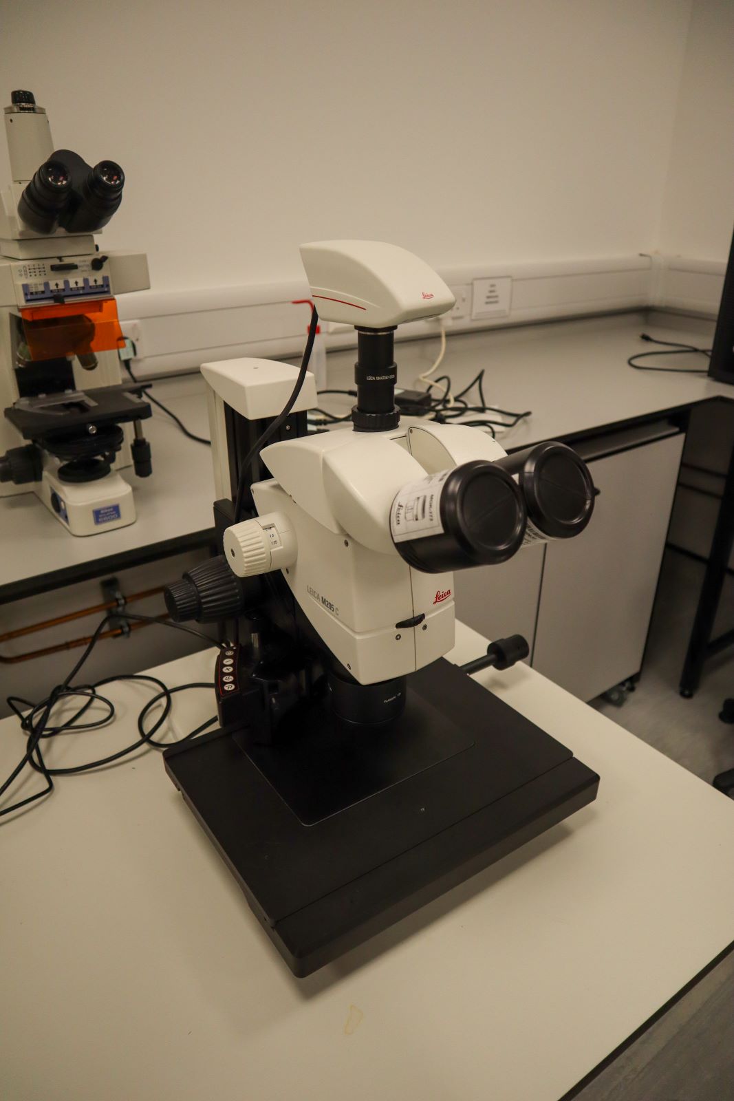
We have a range of digital and optical microscopes and software for the imaging and analysis of a variety of materials.
Please contact us to discuss your needs. Research collaborations are welcome.
Scanning Electron Microscopy (SEM) is capable of extremely high magnifications far beyond what can be achieved with a traditional light microscope. All types of dry sample can be imaged, and gold/carbon coating is available for specimens that are not electrically conductive. In addition to high magnification imaging, this SEM is equipped for energy-dispersive X-ray spectroscopy (EDX). This is an analytical technique used for the elemental analysis of a sample and can be used to map the elemental composition of the surface or cross-section of a specimen. Our service can include sample sectioning as well as preparation (e.g. fixation, dehydration, mounting and coating).
We have one inverted and three upright epifluorescent microscopes available for quick and easy imaging of fluorescent samples. These are all equipped with digital cameras and user-friendly image processing software for quantitative analysis. Our inverted fluorescent microscope is part of our tissue culture suite and is dedicated only for the imaging of live cells with GFP/YFP. The upright microscopes can be used for UV, blue and green excitation (i.e. blue, green and red fluorescence).
FTIR (Fourier transform infrared) spectroscopy helps to identify unknown materials, chemicals or coatings. It may confirm reaction products and help in measuring progress of a reaction. Our machine is also combined with an infrared microscope which can provide high magnification infrared images allowing for further detailed analysis. This can help fingerprint organic functional groups present on localised locations within a sample.
We have a range of digital and optical microscopes and software for the imaging and analysis of a variety of materials.
We are pleased to offer a custom digital slide scanning service. Our Epredia Pannoramic MIDI ll slide scanner and high-quality camera has a throughput of 15 slides per hour with continuous loading for imaging up to 90x in brightfield. Features include automatic loading, previewing and barcode reading. Highest throughput is 15 slides per hour.
Our CytoFLEX flow cytometry platform is equipped with three active lasers (violet [405 nm], blue [488 nm] and red [638 nm]) and nine channels for fluorescent detection across 13 repositionable bandpass filters. Training packages and access to cell culture facilities and expert FlowJo analysis software are available.
Terms and Conditions
Please contact us for pricing and full terms and conditions: NAIF@northampton.ac.uk
Contact us to discuss your needs, our services range from providing access only to full-service solutions. We offer custom training packages, support with experimental design, sample preparation and data and/or statistical analysis. Email NAIF@northampton.ac.uk