Bioscience Laboratories
Our Bioscience laboratories are modern and exceptionally well equipped with research facilities that will enable you to learn and develop your skills in an innovative and supportive environment.
Specialist facilities include:
- the Creative Hub Teaching Laboratory, where students learn fundamental techniques using equipment such as fluorescence and micropipettes, as well as bright-field and electron microscopes. Here students work with real samples such as DNA, RNA and cholesterol to build the foundations of their laboratory learning
- dedicated cell culture facilities, where students work on in-depth studies looking at cancer types, Duchenes muscular dystrophy and sepsis
- specialist microbiology laboratories, used for detailed investigations of yeast, bacteria and fungi, looking into sub-lethal doses of antibiotics and resistance
- chemistry facilities, where we utilise acids and bases, and perform enzyme experiments
- molecular laboratory facilities, used in advanced studies that may involve PCR (Polymerase Chain Reaction), gel electrophoresis, next-generation sequencing technology and western blots, a laboratory technique used to detect a specific protein in a blood or tissue sample
- the Northampton Advanced Imaging Facility (NAIF), which is the University of Northampton’s core facility for advanced imaging and flow cytometry. A wide range of equipment, training and services are available, including:
- Scanning Electron Microscopy (SEM) is equipped with energy-dispersive X-ray spectroscopy (EDX), which provides high-resolution imaging of a sample’s surface while simultaneously identifying and mapping its elemental composition.
- Epifluorescence microscopy allows visualisation of specific structures or molecules within a sample.
- FTIR (Fourier transform infrared) spectroscopy enables the identification (and optional spatial mapping) of chemical compounds in sample based on their characteristic infrared absorption spectra.
- Slide scanning creates high-resolution digital images of entire microscope slides, enabled detailed visualisation, analysis and sharing.
- Flow cytometry allows rapid, quantitative analysis of the physical and chemical characteristics of individual cells or particles as they flow through in a fluid stream through a laser.
Bioscience lab gallery
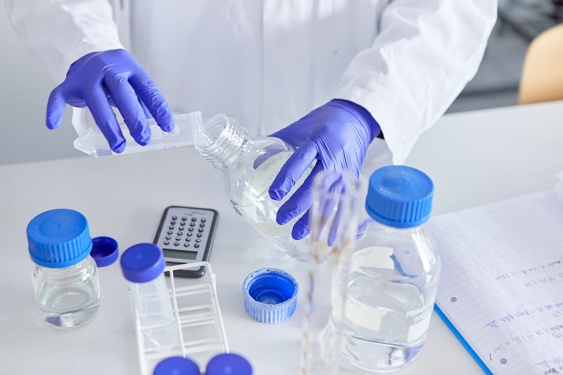
Photo shows a pair of hands wearing purple gloves which are pouring liquid into a container during an experiment in UON's teaching lab
- Courses: Biochemistry BSc, Biological Sciences BSc, Biomedical Science BSc, Molecular Medicine MSc, Pharmacology BSc
- Location: Creative Hub